
Treacher Collins Sydrome
Contents
Overview
In 1900 a British ophthalmologist described two patients with this condition, and as a result this syndrome now bears his name. Treacher Collins syndrome has been shown to occur with the same frequency in boys and girls. Currently, no one knows what causes this condition to occur. In over 60% of cases both the mother and father of a child born with Treacher Collins syndrome have normal genes, and the mother does “everything right” during her pregnancy. The process of bringing genes together from a mother and father is quite complicated. Once in awhile, a gene can be changed in the process. If this occurs it the “right” gene, Treacher Collins syndrome results. This gene has been identified and is sometimes called the “Treacle gene.” This gene is located on chromosome 5q. The chances of having a child with Treacher Collins syndrome are about 1 in 10,000 births. Treacher Collins syndrome can be inherited, and is transmitted in what geneticists call an “autosomal dominant pattern.” This means that if a child affected with Treacher Collins syndrome decides to have children when he or she is grown, there is a 50% chance of having a baby who also has Treacher Collins syndrome. In the very near future, it will be possible for individuals who have this syndrome, to elect to not pass on this trait. However, in order to do this, it will be necessary to undergo in-vitro fertilization and select embryos that do not have the gene for implantation.
Physical Traits

Treacher Collins syndrome presents with different severities. That is, sometimes the syndrome is so mild that it is hard to tell if a child even has the syndrome. Other times, it can be quite severe. The following is a list of traits that a child may, or may not have.
- A narrow forehead.
- Eyes that tilts downward (called an “antimongoloid” slant).
- Pulled down lower eyelids (sometimes erroneously called a “coloboma”).
- Absent eyelashes on the lower eyelids.
- Thin skin overlying absent cheekbones (orbital clefts with absent zygomas).
- Absent ears (microtia), or malformed ears.
- Cleft palate.
- Small lower jaw
Some children may be born without a soft palate (back part of the roof of the mouth) and small, or absent thumbs. These children may have what is called Nager variant.
Treatment
The treatment of a child born with Treacher Collins syndrome (TCS) is complex, and is probably best provided by comprehensive craniofacial teams at major centers. The following is a brief overview of our recommended treatment protocols. Specifics should be discussed with your team of doctors.
Treatment recommendations are constantly changing over time, and will also vary from center to center. It is important to discuss these issues with your doctor, and make sure that all your questions get answered.
The First Year of Life
After a child is born with Treacher Collins syndrome, the child should undergo a hearing screening as soon as possible. Children born with absent ears typically have at least a 40% hearing loss. For these children, it is very important for them to be evaluated early for a bony conductive hearing aid. We now know that the early sounds babies hear, are critical for normal speech development.
It is also important to evaluate the palate, and assess palatal function. Some children who do not have an obvious palatal cleft may still have what is called a “submucous cleft.” This is a condition where the muscles normally found in the roof of the mouth are oriented in a different way. These children need to be closely followed by a speech pathologist, for sometimes they may require surgery to enable them to speak normally.
Treating Sleep Apnea
It is also important to make sure that any child with Treacher Collins does not have sleep apnea. If your child is a noisy breather at night, it is a good idea to get a sleep study to ensure your child is getting enough oxygen at night to continue to develop normally. For some children, it is possible to use a special orthodontic device (a “Herbst appliance”) to help prevent sleep apnea. Rarely, children may need to use a special mask (called CPAP or BiPAP units), in order to help the child get enough oxygen while sleeping. If these do not work, surgery may be the only other option to ensure your child is getting enough oxygen at night (see The Jaws).
The Forehead
Only a small percentage of children with TCS require surgery on their foreheads. Typically, this surgery is only necessary in a few of the most severely affected children. This operation normalizes the shape of the forehead and eyebrow bones.
The Eyes
The inferior tilt of the outside of the eyelids (an area called the lateral canthus) is best treated with an operation called a “canthopexy.” This operation basically just lifts the outside of the corner of the eyes. In general, people are considered more attractive if the outside corner of their eyes is higher than the inside. In TCS, the eyes tilt downwards. By lifting up the outside corners, the child will appear to look “less sad.” This operation can be done at any age, however, the younger the child is, in general, the less successful this operation is because the eyes will tend to go back to the way they were. This recurrence is because of a substance in the skin that is called elastin. Elastin works like a rubber band, it helps to keep youthful skin tight, but also “wants” to pull the corner of the eye back to where it was before surgery. As we all get older, we lose this elastin, and as a result the effects of surgery are more dramatic. At our center, this operation (a canthopexy) is typically done without putting any external facial scars on the patient.
Another operation that may be recommended is the upper eyelid switch flap. In this operation, tissues are taken from the upper eyelid and brought down to the lower eyelid. I almost never recommend this operation. The scars that are left behind (in my opinion) tend to look horrible for the rest of the child’s life. Most patients that have had this procedure are very unhappy with the results, yet many doctors still recommend it to their patients. The use of skin grafts in the lower eyelids also look very bad, and tend to be ineffective, as well.
The Ears
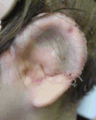
For children born with absent ears (microtia), there are two options to rebuild ears, one is to get artificial ears, and the other is to rebuild the ears using the child’s own rib (also called an autogenous reconstruction). I believe that it is important that the child participate in the decision as to which technique would be best. The pro and cons of each method of reconstruction are discussed in an office visit. For those families choosing an autogenous reconstruction, this series of operations typically begins sometime after the child has turned eight-years old. The best time to rebuild the ears is determined by the child’s size and how the child is coping socially. Children with TCS are typically smaller than their classmates, and it is necessary for children to be a certain size before ears can be made. Ears are reconstructed over a series of three operations. Some doctors believe that both ears should not be built at the same time. However, in Dallas we have had good success with building both ears at the same time, and believe that this is the best way because it reduces the total number of operations a child must undergo.


The first stage ear reconstruction is shown above. Cartilage is taken from the child’s rib (parents cannot donate their rib cartilage) and this cartilage is carved and assembled to create the ear. It is then inserted under the skin. Two additional stages are required to complete the reconstruction; both performed as outpatient procedures. Children with TCS are more likely to require hair removal, as can be seen below, in a child who has just finished the second stage procedure. The third stage will involve making a hole that looks like an ear canal.
Before undergoing ear reconstruction, it is wise to ask your doctor to see examples of his or her work. It is very difficult to rebuild an ear that did not turn out well. It is also possible for children to undergo reconstruction of the absent eardrum (by a specialized ENT), in order to try to improve hearing. The decision as to whether or not to proceed with inner ear reconstruction should be made with a specialist in this area. Our office would be happy to make some recommendations as to who to see. Typically, this procedure is not possible for most patients with TCS, because the inner ear may not be formed well enough to give a good result. It is best to wait until the child is about 9-years old before undergoing the specialized CT scan that will determine whether, or not, it is possible to create an eardrum.
Cheekbones
The cheekbones are usually 80-90% of their adult size by the time a child reaches 9-years old. Therefore, it is wise to wait until at least this old before trying to reconstruct this area. I prefer to rebuild the missing bone, with the child’s own bone. The bone to rebuild this area is usually taken from the skull for two different reasons. The first is that bone taken from the skull is less likely to disappear after the operation (bone taken from the rib, or hip, is more likely to dissolve away). The second reason is that there is almost no pain associated with taking bone from the skull. Is taking bone from the skull safe? In Dallas, we have been fortunate to have not had any complications from this technique. The safety of this technique, when performed at our center, has been published (see publications #16), but it is certainly possible for a patient in the future to have a problem. In addition to rebuilding the absent cheekbones, often children will undergo a transfer of underlying soft tissue to the cheeks, a reshaping of the nose, and a lifting of the corners of the eyes, all in the same operation.
When the face has finished growing (around age 17), and all necessary jaw surgery has been completed, then it is safe to place artificial cheekbones to complete the reconstruction. This gives the child their final, normal-looking, adult sized cheekbones. It is very important that these implants not be placed too young, while the child is still growing, because they will sink too deeply into the underlying bone. More importantly, if any upper jaw surgery is performed after the cheek implants are placed, they will have to be removed in order to prevent a serious infection.
The Jaws
Children with TCS almost always require both braces and jaw surgery. Typically, children’s upper jaw grows too far forward compared to the lower jaw. However, sometimes the reverse may occur. It is also fairly typical that the front teeth cannot close together. That is, the back teeth may hit, but up front there is a gap between the teeth. This condition is called an “open bite” and it can only be treated surgically, by operating both on the upper and lower jaws. It is a condition that is very difficult to treat, and may take more than one operation to get just right.
Some doctors recommend using a technique called “distraction” on the lower jaw in order to stretch it forward. This technique is not used at our center for a number of reasons (listed below). This technique involves making an osteotomy (cut in the bone) across the mandible (lower jaw) and attaching a metallic screw driven device to either side of this osteotomy. Pins are usually put through the skin into the bone, and the expansion device is attached to these pins. By turning a screw on the expansion device, a little each day, the bone slowly gets longer. There are also devices are completely buried under the skin, except for a small metal bar that pokes outside so that the screw can be turned. These distraction devices successfully lengthen bone, and the majority of doctors treating Treacher Collins syndrome recommend them. However, these devices are almost never used at the Craniofacial Center in Dallas. There is a long list of reasons why I chose not to use these devices for this condition:
- The overall complication rate for using these devices is about 1 out of three cases, as published in a recently survey of doctors. The complication rate was over 50% for doctors who had done fewer than 10 patients! Surveys typically underestimate complication rates, so that the actual complication rate may even be higher.
- These devices leave scars on the child’s face that cannot be removed, and remain for a lifetime. Many doctors say that the scars will fade away and not be noticeable. I recommend seeing another child’s scars first (in person, scars tend to not show up in photographs) before allowing this procedure to be done. There is an internal distraction device that does not leave facial scars, but this device has an even higher complication rate than the external device.
- In order to attach the distracter to the jaw, it is necessary to use metal pins or screws. It is very likely that the front pins or screws will go through the child’s un-erupted permanent teeth. The result is that the permanent tooth will not come up and the child will need an artificial tooth.
- The upper jaw is not treated by distraction. This means that after the two operations are finished, one to put on the distracter and one to take it off, the upper jaw is still out of position, and will need surgery to correct it.
The alternative to using distraction devices is a single-staged orthognathic procedure. From inside the mouth (so there are no scars on the face) the upper and lower jaws may be cut and moved into a better position. It is best to delay this surgery until the child is older and wants to have the surgery done, unless the jaws are way out of alignment or the child has a tracheostomy. This operation is longer and more complicated than putting on a distracter, and not every doctor is able to do this procedure; but the final result is usually better. However, in very severe cases, in which the jaw is extremely small, there may be an indication for a new type of distraction.