
Microtia
Contents
Overview

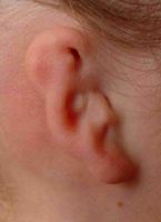
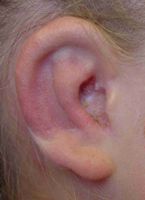
Microtia literally means “small ear.” This term is applied to anyone who has a smaller, or absent ear (although technically speaking, the absence of any ear remnant is referred to as “anotia”).
Microtia occurs in approximately 1:6000 births. For some reason (currently unknown), microtia occurs more often on the right side than the left. This condition can also affect both ears.
Any child born with microtia should be evaluated at a craniofacial center to rule out other conditions. For example, children born with microtia on one side may have a condition called Hemifacial Microsomia (see the section on Hemifacial Microsomia). Children who have both ears affected might have Treacher Collins syndrome or Bifacial Microsomia.
What causes Microtia?
At the current time, no specific gene has been identified that causes this condition, and almost never does someone with hemifacial microsomia pass the trait on to his or her children (less that 5% chance). The exact cause of this condition remains unknown but there is some experimental evidence, published many years ago, that suggested if a small blood vessel ruptures near the developing ear in mice before they are born (so this area ends up getting less blood), after birth the mice will have an absent ear. More likely, this condition results from an impaired flow of cells (called neural crest cells), which arise adjacent to the spinal cord and travel to the face to form the facial skeleton. We know that if not enough of these cells are able to successfully migrate to their intended location, the ear is either smaller, or absent. It has been suggested that some medications may lead to this condition, but this is extremely hard to prove. In conclusion, all current evidence suggests that many complicated factors lead to microtia, and there is nothing that the mother did wrong during pregnancy to cause this to happen.
Can my child hear out of this ear?
When children are born without an external ear, or ear canal, usually the inner one half of the ear is normally formed. This means that children can hear even though the outer ear is not present. If you stick your fingers in both ears, you can still hear. This is approximately how much a child can hear out of the affected side. Most children lose about 40-50% of their hearing if they don’t have an ear canal with an eardrum. However, children seem to hear just fine because the other ear is not affected. The only problem that children with microtia may have is in localizing sound. Without the ability to hear in “stereo,” children cannot tell where a sound, or someone calling their name, is without being able to see them.
Treatment
Early
When a child is born with an absent ear, keeping the opposite functional eardrum healthy takes on an even greater importance. I recommend that parents who have a child with microtia be on the lookout for any sign of an ear infection on the opposite side (it is not really possible to get an ear infection on the microtic side). Multiple ear infections on the unaffected side may diminish hearing; therefore, parents should have a low threshold for bringing their baby to the pediatrician (or pediatric Ear, Nose and Throat specialist) to check for possible ear infections. In general, the vast majority of children with microtia never need any hearing aids. It is important to have your child’s hearing tested periodically to ensure the health of the unaffected ear.
Late
There are two options to rebuild the ear, one is to get an artificial ear, and the other is to rebuild the ear using the child’s own rib (also called an autogenous reconstruction). I believe that it is important that the child participate in the decision as to which to ear would be best. The pro and cons of each method of reconstruction are discussed in an office visit. For those families choosing an autogenous reconstruction, the outer ear is typically reconstructed around age eight, but on occasion may be done as early as six. The best time to rebuild the ear is determined by the child’s size (more importantly, how big the child’s ribs are) and how the child is coping socially. Ears are reconstructed over a series of three operations.


The first stage ear reconstruction, in a child with hemifacial microsomia is shown above (upper row, left). Cartilage is taken from the child’s rib (parents cannot donate their rib cartilage) and this cartilage is carved and assembled to match the opposite ear (upper row, right). It is then inserted under the skin. I do not place any bandages for the first stage, although most doctors do wrap up the entire head with a bandage; I have learned that these bandages do not do anything to help the child, and only make the operation more difficult for the child. Two additional stages are required to complete the reconstruction; both performed as outpatient procedures. The second stage involves cutting behind the ear and lining this space with a skin graft. After surgery I cover this new skin with a yellow bandage (bottom row, left) that is removed about a week later. The appearance of the ear after the second stage is seen in the photograph on the bottom row, middle. The third operation creates the appearance of an ear canal, but it does not help hearing. Following this third, and smallest procedure, the ear is finished (bottom row, right).
There are a number of variations on the techniques used to rebuild an ear, and different doctors may use different techniques. Many people may have seen a picture of an experimental mouse with an ear growing on it’s back. Unfortunately, attempts to transfer these grown ears to the head have ended up looking terrible. The experience and the artistic ability of the doctor performing the operations are probably more important than the specific technique the doctor chooses. Before undergoing ear reconstruction, it is wise to ask your doctor to see examples of his or her work. It is very difficult to impossible to rebuild an ear that did not turn out well the first time.
Following outer ear reconstruction, it is also possible for children to undergo inner ear reconstruction (by a specialized ENT) in order to rebuild the absent eardrum. This is done to try to improve hearing on the affected side, and achieve some stereo hearing that improves the localization of sound. This operation is not without some downsides that need to be discussed with your doctor. I also strongly recommend speaking with other patients who have undergone inner ear reconstruction before deciding to proceed with inner ear surgery. For those who decide to proceed with inner ear surgery, it is important to first finish building the external, or outer ear.